


| Homo sapiens |
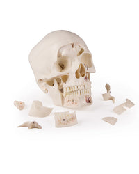
Replica Luxury Demonstration Skull, 14-part
Regular price
CHF 596.00
Category for species identification
Item Description
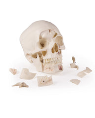
This 14-part skull model is a cast of a real human skull specimen and displays all anatomical structures in the highest detail. It was developed for students of anatomy, medicine, surgery, ENT, ophthalmology, and dentistry. The skull is intricately cut and assembled with metal and magnetic connections.
The cranial vault can be opened and removed, leaving the temporal bones and their sutures untouched. Bony impressions of the sagittal sinus, transverse sinus, and sigmoid sinus, as well as the meningeal vessels, are painted. The skull base is sagittally cut in such a way that the cut on one side passes through a cribriform plate and another cut on the same plane passes through the other cribriform plate of the ethmoid bone, leaving the crista galli and perpendicular plate, as well as the entire nasal septum, intact. The structures of the anterior, middle, and posterior cranial fossae are easily accessible. One can directly see the nasal cavity, nasal concha, septum, as well as the bony pharynx and nasopharynx. The nasal septum can be removed from the surrounding bone structures. The frontal sinuses are prepared as a whole on one side and opened on the other side, making them fully accessible. The relationship of this sinus to the nasal cavity is clearly visible, which is particularly valuable for ENT doctors.
On one side of the skull, the temporal bone has been left in situ. The other temporal bone is removable from the skull. A part of the mastoid process and the squamous part of the temporal bone, along with the mastoid antrum, can be removed, offering a clear view of the inner ear. All three semicircular canals are visible along with the course of the facial nerve, which runs posteriorly and then inferiorly, ultimately exiting through the stylomastoid foramen. The removable temporal bone shows a complete external auditory canal. An almost vertical cut through the mastoid process and then inwards along the petrosquamous fissure divides the temporal bone, and the position of the eardrum can be seen. The carotid canal is opened, as is the cochlea, showing the internal auditory canal, and the course of the facial nerve can be observed. The oval window, the semicircular canals, and the opening of the tympanic cavity are visible.
The maxilla and mandible show the structures of the dentition, the roots, the bony edge of the alveolar process, as well as dental nerves and vessels. The maxillary sinus can be opened by removing a flap. The teeth of the right mandibular flap are bisected, showing the internal structures of the teeth.
Article number
Artikelnummer: 4800
Barcode: 4250395772331
Shipping Information

PostPac Economy
ab CHF 9.50
PostPac Priority
ab CHF 11.90
YouTube
Derzeit kein Video verfügbar.
Payment methods


Replica Luxury Demonstration Skull, 14-part
Regular price
CHF 596.00
ERLER ZIMMER
Replica Luxury Demonstration Skull, 14-part
Werbung
